Test info

Electromyography/Nerve Conduction Test and Neuromuscular Ultrasound Explanation

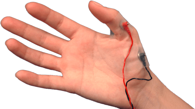
EMG and NCS tests are done in order to evaluate the condition of nerves and muscles. During the test there may be a very mild electrical stimulation applied to your extremities, and a very thin electrode-needle will be inserted into some of the muscles in order to observe their reactions. The test makes use of highly-sophisticated computerized equipment, which allows calculations of the rate at which your nerves conduct electrical impulses, and it assesses whether or not your muscles may have had some interruption in their nerve supply, or whether there is any damage in the muscles. This gives your doctor information about such disorders as injured nerve roots, damaged nerve and muscle disorders, which will allow a better understanding of your problem and provide for the best possible treatment.

There is often some discomfort during the procedure, but it is usually mild, and you should feel free to ask for an explanation or a rest in between various parts of the study as it progresses. Generally, the complete examination requires an hour.

Please do not use any lotion, ointments, creams, or powder on your skin for 24 hours prior to the test. Clean skin is imperative. However, you may use deodorant.

In certain cases, we may recommend a neuromuscular ultrasound. This is a painless procedure that helps your doctor see nerves, muscles, and soft tissues in real time, giving a clear view of these important structures.

When paired with electromyography (EMG), neuromuscular ultrasound combines detailed images of the body’s structure with information about how these structures are functioning. This combination enhances our ability to diagnose and understand various conditions.

During the procedure, a small amount of ultrasound gel is applied to your skin. Then, an ultrasound probe is gently moved over the area, allowing us to visualize the underlying structures. This process is straightforward and non-invasive, ensuring your comfort while providing us with valuable insights.

Click on the play button to view our EMG Test Video

Haz clic en el botón de Reproducir para ver nuestro video de Prueba de EMG

Explicación De La Prueba De Electromiografía (EMG), Del Examen De Conducción Nerviosa (NCS) Y Ecografia Neuromuscular

El EMG y el NCS son pruebas que se hacen para medir la velocidad con la cual sus nervios conducen el impulso nervioso (similar a la conducción de la electricidad) y para medir la manera como sus músculos responden a cierto estímulo. Durante la prueba se le insertará una aguja muy pequeña en algunos musculos a través de la cual se le aplicará una estímulo eléctrico de muy bajo voltage. Durante el examen, se utiliza un equipo de computación altamente sofisticado, el cual nos permite calcular la velocidad a la cual sus nervios conducen el impulso nervioso y determinar si existe algun daño en el músculo. Esta información ayudara a su médico a diagnosticar problemas, incluyendo lesiones en la raiz del nervio u otras abnormalidades tanto en los nervios como en los músculos, y de esta manera entender su problema y ofrecerle el mejor tratamiento posible.

Usualmente, los pacientes sienten alguna molestia durante el examen, pero ésta es generalmente leve.
Usted puede pedir que se le explique los que se le está haciendo durante el examen,
e incluso que lo interrumpamos momentaneamente. El examen tiene una duración de 1 hora.

Por favor, no use aceite, loción, crema o talco en la piel 24 horas antes del estudio.
Sólo se permite el uso de desodorante. Limpiar la piel es indispensable.

En ciertos casos, podemos recomendar una ecografía neuromuscular. Este es un procedimiento indoloro que nos permite visualizar los nervios, los músculos y los tejidos blandos en tiempo real, brindándonos una imagen clara de estas estructuras importantes. 
 
 Cuando se combina con la electromiografía (EMG), la ecografía neuromuscular une imágenes detalladas de la estructura del cuerpo con información sobre el funcionamiento de estas estructuras. Esta combinación mejora nuestra capacidad para diagnosticar y comprender diversas afecciones. 
 
Durante el procedimiento, se aplica una pequeña cantidad de gel de ultrasonido sobre la piel. Luego, se desliza suavemente un transductor de ultrasonido sobre el área, lo que nos permite visualizar las estructuras subyacentes. Este proceso es sencillo y no invasivo, garantizando su comodidad mientras nos proporciona información valiosa.

Medical illustrations on this website are attributed to: Freepik.com and author Kjpargeter

Lateral Femoral Cutaneous Nerve

From: www.wheelessonline.com 

– Anatomy:    

– LFCN, as its name suggests, is purely sensory;    
– it arises from L2 and L3, travels downward lateral to the psoas muscle, crosses the iliacus muscle (deep to fascia), passes either thru or underneath the lateral aspect of the inguinal ligament, and finally travels onto innervate the lateral thigh;
– it divides into anterior and posterior branches and supplies skin on lateral aspect of thigh;
– in the study by Hospodar et al (JTO 1999), the course of the nerve was variable, but was most commonly found at 10-15 mm from the ASIS and as far medially as 46 mm from the ASIS;
– in no specimen did the nerve pass lateral to the ASIS (eventhough historically the nerve is thought to pass lateral to the ASIS in 10% of population);
– in all specimens the nerve passed underneath the ilioginal ligament and anterior to the iliacus muscle; 

– Meralgia Paresthetica: 

– entrapment syndrome of the lateral femoral cutaneous nerve causing burning, numbness, and paresthesias down the proximal-lateral aspect of the thigh;
– may be idiopathic, be a result of trauma, previous operations, and in some cases may arise from Perthes Disease abduction splints;
– in idiopathic cases, the nerve may be encased in bone by the growing apophysis of the anterior superior iliac spine, or may be entrapped in fascia either proximal or distal to the ASIS;    
– diagnosis is made by:           
– reproduction of the pain by deep palpation just below the anterior superior iliac spine and by hip extension;           
– relief of pain by localized injection of lidocaine;    
– treatment: when diagnosis is not in doubt and the symptoms are severe, consider operative decompression at the site of constriction;

EMG and NCS

Electromyography (EMG) and nerve conduction studies (NCS) are tests that measure the electrical activity of muscles and nerves. Nerves send out electrical signals to make your muscles react in certain ways. Nerves also send signals from your skin to your brain, which then processes the signals, so you can experience a variety of skin sensations.

  • An EMG Test looks at the electrical signals your muscles make when they are at rest and when they are being used.
  • A Nerve Conduction Study measures how fast and how well the body’s electrical signals travel along your nerves. EMG and nerve conduction studies are used to help diagnose a variety of muscle and nerve disorders and to quantify the severity of your condition.


An EMG test helps find out if muscles are responding the right way to nerve signals. Nerve conduction studies help diagnose nerve damage or disease. When EMG tests and nerve conduction studies are done together, they help doctors tell if your symptoms are caused by a muscle disorder or a nerve problem. With EMG and NCS studies, your doctor can next select the best therapeutic option to help you.