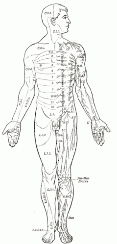
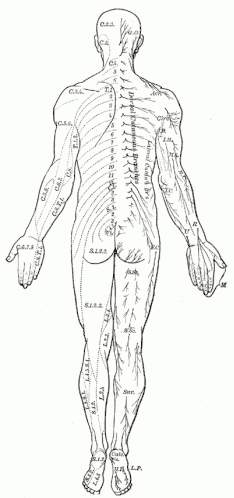
Radiculopathies
Radiculopathies are very frequently encountered in clinical practice. The most common deferential diagnosis includes:
Non-Neuromuscular (orthopedic) Lesions:
- Muscle strain/sprain
- Rotator cuff tendonitis/tear
- Subacromial/trochanteric bursitis
- Epicondylitis
- Degenerative joint disease
Neuromuscular Lesions:
- Motor neuron disease
- Brachial and Lumbosacral Plexopathy
- Mononeuropathy
- Polyneuropathy
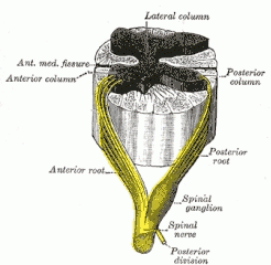
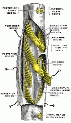
Cervical Radiculopathies:
- C5: 2 – 14%
- C6: 19 – 25%
- C7: 56 – 70%
- C8: 4 – 12%
Lumbar Radiculopathies:
- L2/3
- L4
- L5 : most common
- S1: second to L5
The goal of the EMG is to localize the compressed, and to establish the presence or absence of active axonal loss and/or chronic reinnervation changes. A very important role of the NCS/EMG is to evaluate for presence of a “Double Crush Syndrome”, in which a distal mononeuropathy is superimposed. The electrophysiologic abnormalities in radiculopathies include:
Possible abnormalities in motor NCS:
- Normal sensory NCS (lesion is proximal to DRG)
- Possible active denervation in appropriate myotome and paraspinals
- Very important limitations of EMG in radiculopathies are:
- Absence of abnormalities in acute lesions (< 2 weeks)
- “Normal” EMG in purely demyelinating lesions (no denervation)
- “Normal” EMG in lesions limited to the sensory nerve root